
The Importance of Ultrasound
If your doctor has requested an Ultrasound Scan, you may be feeling a little apprehensive or curious about the procedure.
The good news is that ultrasound scans are safe and pain-free in the hands of an experienced and highly Specialised Radiologist Doctors.
There is no radiation involved, and in most cases, the exam is entirely non-invasive.
What is an ultrasound?
An ultrasound is a safe, generally non-invasive medical imaging and diagnostic tool. Other names for ultrasound include sonogram and ultrasonography.
Our private pregnancy ultrasound scan services use this safe technology throughout your pregnancy journey.
Ultrasounds use sound waves to generate images of the inside of your body, allowing radiologists and your doctor to see your organs, tissue, and other structures.
They do not use radiation like X-rays and other medical imaging technology. Broadly, ultrasounds are used for one of two reasons:
Pregnancy ultrasounds examine an unborn baby and check its health and growth status.
Diagnostic Ultrasounds offer information about internal areas and parts of the body, such as the liver, kidneys health, and reproductive organs.
While they are a powerful and valuable tool in the right setting and under the guidance of experienced, qualified specialists, there are shortcomings.
Ultrasounds cannot look at every structure in your body. For example, because sound waves don’t travel well through bone, ultrasound is not used to look at the brain and other organs covered by bone.
How ultrasound imaging works
Ultrasound uses sound waves to produce black-and-white images of the body’s internal structures. Waves are produced by a wand-like device called a transducer, which typically uses ceramic crystal materials called piezoelectrics.
Similar principles apply in early pregnancy scans, where we capture images of your developing baby.
Piezoelectrics create sound waves when an electric field is applied and create an electric field when sound waves are applied.
This allows the transducer to detect waves that bounce off boundaries between structures in your body — for example, the boundary between tissue and bone.
The scanner measures the speed and timing of each reflected sound wave. It uses this data to determine the distance between the transducer and the boundary. These distances are what generate images.
The value of ultrasounds as an imaging and diagnostic tool
Ultrasounds are a valuable imaging tool in the hands of a radiologist because they pose zero risk, are non-invasive, and can be used to examine many areas of the body.
A pregnancy ultrasound is generally undertaken to examine the health of the baby. If you are pregnant, you might have a scan to determine the following:
The size and position of your baby
How long you have been pregnant for (gestational age)
If you are pregnant with multiples
If your baby has congenital disabilities in the spinal cord, heart, brain, and other areas of the body
The volume of amniotic fluid, which surrounds and protects unborn babies
Diagnostic ultrasound can give your doctor information about the following:
Your Liver and gallbladder’s health
Cancerous or non-cancerous lumps in your thyroid gland
Abnormalities in your kidney and abdomen
A breast lump For women specifically, diagnostic ultrasound may be used to:
Uncover the source of pelvic pain
Diagnose infertility
Determine the cause of abnormal menstrual bleeding
While a unique and widely used tool, it does have limitations. For example, it cannot produce images of every area in the body, particularly those covered by bone or that contain gas.
Your doctor may request an X-ray, CT scan, or MRI to examine these areas.
What to expect from the ultrasound process
Ultrasounds are not painful, require minimal or no preparation, and enable you to go about your regular day-to-day activities immediately after your scan.
While the examination process will depend on the area of the body being scanned, it generally unfolds as follows:
Your doctor will ask you to lie down on the examination table. You will need to expose the area being examined — that may mean removing some clothing.
Your doctor will apply a special ultrasound gel over the target area. It will likely feel cold.
Your doctor will move a wand-like device, the transducer, over the area. This sends harmless sound waves into your body. The waves are so high-pitched you won’t be able to hear them.
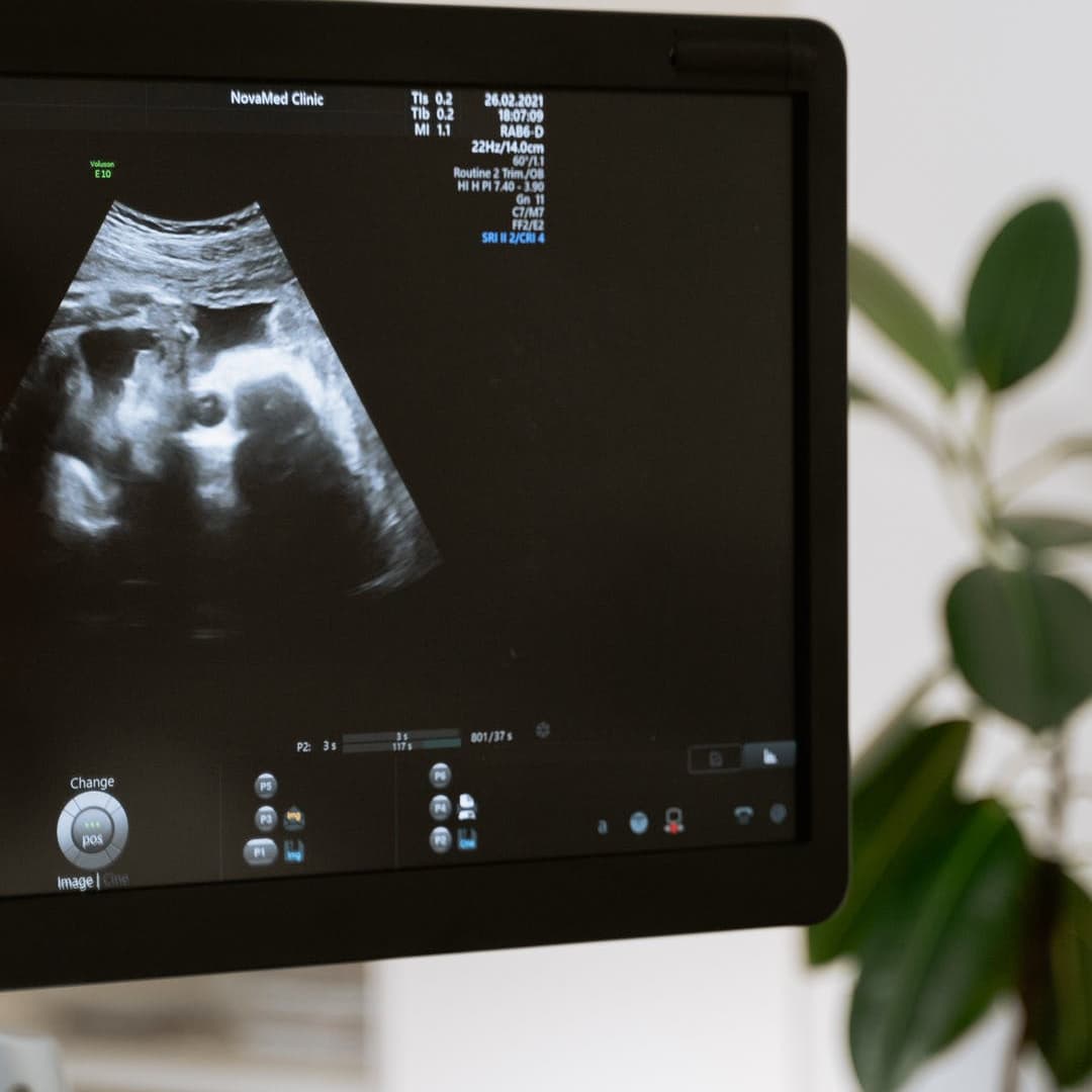
The scanner records the reflected waves and translates them into black-and-white, two-dimensional images on a screen.
You will see the images live on a large screen in front of you. Especially during a pregnancy ultrasound, allowing you to see your unborn baby.
The test will take about 20 to 40 minutes from start to finish. When your doctor has all the images required, they will wipe the gel off the target area and give you privacy to redress.
Reasons why you may need an ultrasound
There are many different reasons why you might undergo an private ultrasound scan. It allows your healthcare provider to check on your baby’s well-being without using any harmful radiation.
You may need a diagnostic scan if you are experiencing symptoms in specific organs or areas. For example, a diagnostic ultrasound can give your doctor information about your reproductive system, gallbladder, thyroid, liver, and kidneys.
For expectant parents wanting to know their baby's sex, our gender scan service is typically performed between 18 and 22 weeks.
You may also need one if you have adverse breast implant symptoms.
Frequently asked questions
How do I prepare for an ultrasound?
There is very little preparation needed. In most cases, you can show up, remove any jewellery or clothing obscuring the target area, and undertake your exam.
However, some scans require special preparation:
For an abdominal scan, you will need to fast for 4 hours before the scan, but you can have water, black tea or coffee without milk or sugar.
For a pelvic exam, you may have to drink a lot of water to fill your bladder so that images can be as clear as possible.
Your doctor will inform you of any specific preparation required if needed.
How long does an ultrasound scan take?
Ultrasound scans can take as little as 20 minutes and as long as one hour — it depends on the area or areas being examined.
Does an ultrasound scan hurt?
No. Ultrasounds are not painful. However, if you have a sore lump to the touch, you may experience some discomfort during the scan.
You may also have to lie in a particular position, which may not be ideal. If you feel pain at any time during your scan, it’s best to tell the healthcare professional performing the ultrasound.
What areas of the body can an ultrasound examine?
The areas of the body an ultrasound can examine are the following:
The abdomen, which includes the liver & gallbladder, spleen, kidneys , pancreas, kidney and urinary tract, and larger blood vessels
The pelvic region, including elements of the male and female reproductive systems
Musculoskeletal structures, including areas like the hip, elbow, or shoulder and knees
Book a Scan Online
Your health matters. If your doctor has requested ultrasound or you are experiencing symptoms you’d like diagnosed, schedule your private ultrasound appointment with Our Specialists at Harley Street Ultrasound.
Our dedicated team is here to make your experience as comfortable and positive as possible. Plus, we offer same-day results accessible via your computer or smartphone.